Explore the microscopic world with a powerful and easy-to use microscope for any smartphone!
DIPLE – the POWERFUL microscope for any smartphone
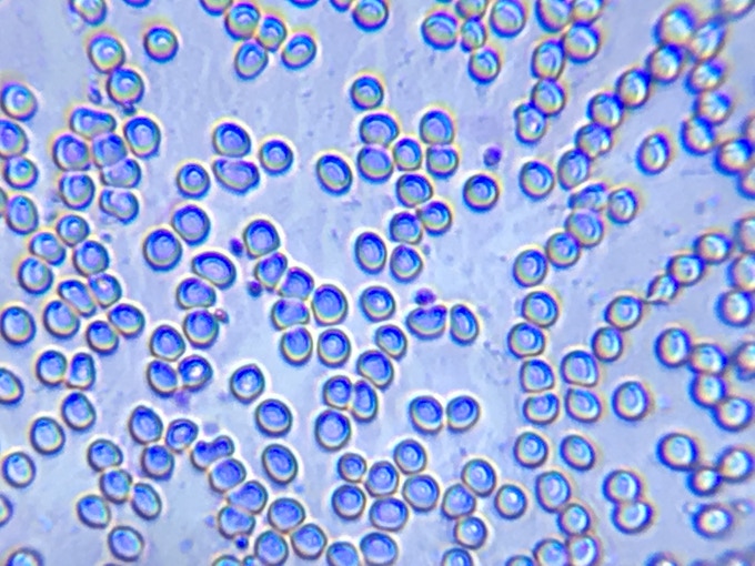
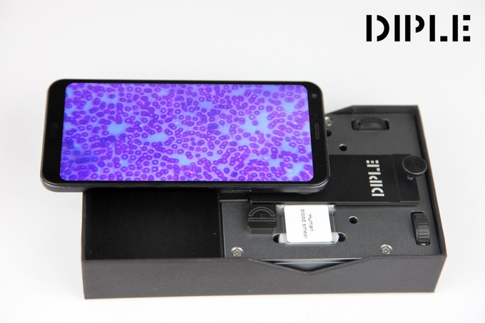
DIPLE for observing human blood cells – Photo courtesy of Istituto Italiano di Tecnologia
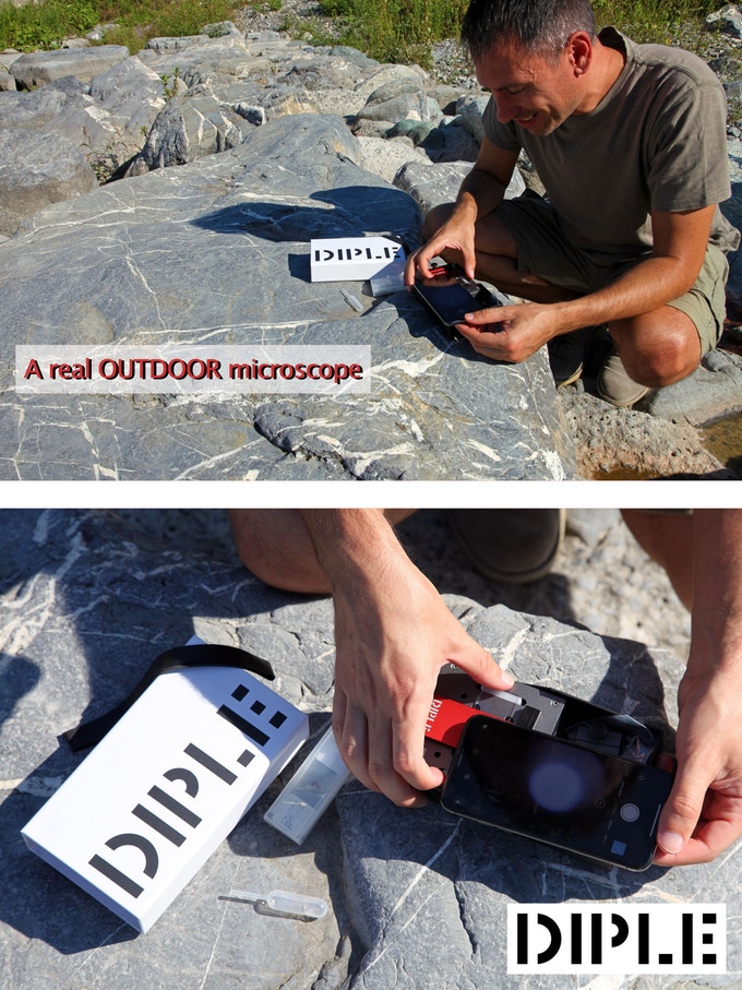
This easy to use, compact, and portable kit transforms any smartphone and tablet into a POWERFUL microscope. Whether you use it for research, education, or just for fun, from the observation of microorganisms or cells to mineralogy and materials science, with DIPLE anyone can access the microworld in detail!
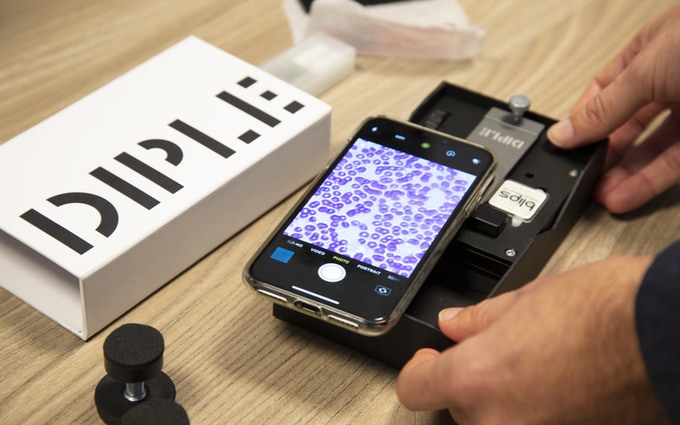
DIPLE is capable of giving you 1000x magnification using your smartphone or tablet as a monitor. Set up takes seconds – all you need to do is place your camera on the optical system. You can even take pictures and videos in HD!
To make sure that DIPLE is truly portable for exploration on the go, we have designed it to easily withstand being dropped – even from the third floor of a building (yes, we did the test!). Available in single or coupled lenses depending on the level of magnification required, such an advanced system has never been used for microscopy on smartphones (it is patent pending). DIPLE provides an unprecedented quality of image for a budget microscope.
The concept
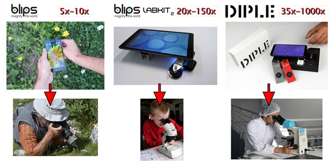
After the great success of the BLIPS kits, our team needed something to get deeper into the microworld. DIPLE is the answer – a completely NEW CONCEPT, to overcome the limits of Blips for microscopic inspection at a smaller scale.
We’ve come back to Kickstarter after three years of development and we can’t wait to bring our community a new view of the world.
Key features
LIGHT, COMPACT, AND PORTABLE: The whole DIPLE system can be taken anywhere. Measuring in at 17,6cm length, 10cm width and 4cm in height, it’s actually smaller than the box your smartphone came in!
EXTREMELY POWERFUL: You can clearly see cellular details, and even bacteria! Thanks to its great optical power, you can digitally zoom the image for achieving an overall MAGNIFICATION OF 1000X before getting any pixelation (with optical resulution below 1 micron).
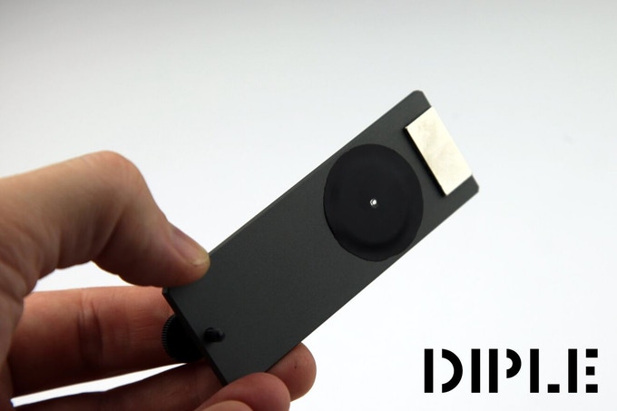
OBJECTIVE LENS: Unlike other products, having the objective lens separate to the phone camera ensures an easier way to find the ideal focal distance, even for EXTRA THIN samples (e.g. blood cells). It also means that it does not depend on the shape of your phone, so it works with all types.
IMMERSION MICROSCOPY: The flat surfaces of the magnifying objective lenses can work when immersed in transparent liquids.
FILTERS: DIPLE can be expanded with filters for specific needs, as its design is derived from the “infinity corrected” microscopes.
MODULAR: This means that you can upgrade your system in the future, getting new single spare parts, or use it for your own optical improvements.
DURABLE: The innovative objective lens of DIPLE is made with metal, meaning the objective lens can drop from 10 meters (33 ft) without issue.
NO FUSS: To keep set up as simple hassle free as possible, you don’t need to remove your phone / tablet case as you would with other products
EXTRA-FINE REGULATION (SUB-MICRON) OF THE FOCAL DISTANCE. A purposefully designed mechanical system allows you to move toward or away from your sample with extreme precision, always obtaining the perfect focus.
ORIGINAL DESIGN: The patent pending design is unique, you do not have to add or clamp anything to the body of your phone.
CARRYING BOX: It’s not just a container, but a functional part of the product. It is the mechanical support for all the components when you are using the product.
AFFORDABLE PRICE: How many portable microscopes allowing you to see bacteria are sold at less than 100USD?

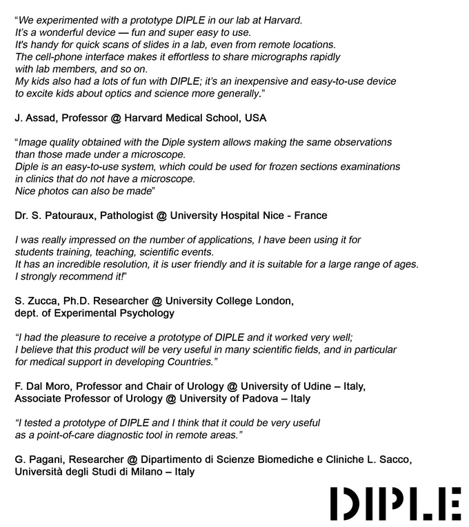
People who love DIPLE
Find your perfect DIPLE
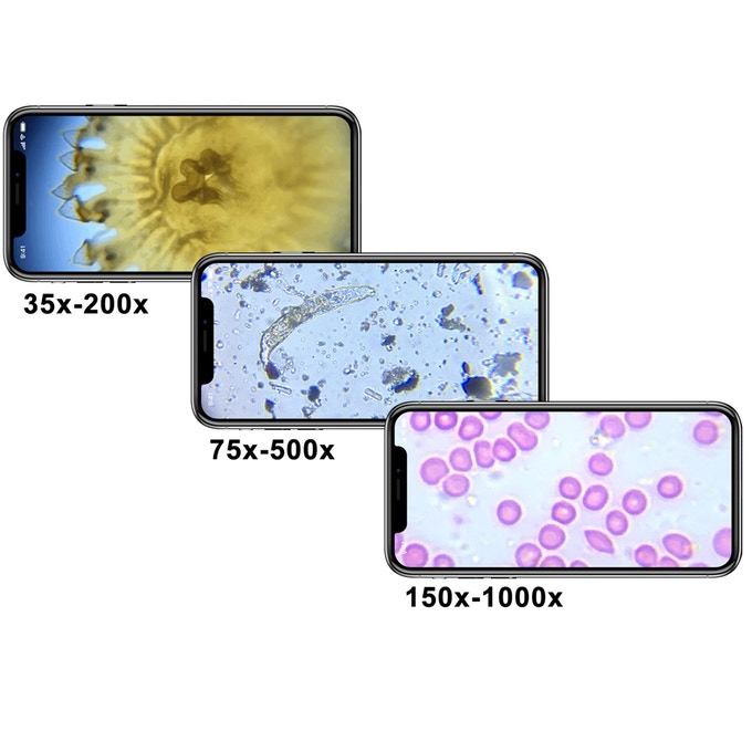
There are three versions of DIPLE objective lenses for microscopy in transmitted light + the new DIPLE LUX for exploring the microscopic details of opaque surfaces.
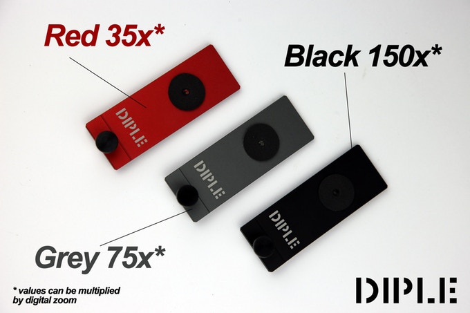
DIPLE RED, providing 35x REAL magnifications – resolution of 3 micron, field of view of around 1 mm – to see cells, microorganisms (protozoans, rotifers, water bears…);
DIPLE GREY, providing 75x REAL magnifications – resolution of 1 micron – to see bacteria, cells, protozoans…
DIPLE BLACK, providing 150x REAL magnifications – resolution of 0,7/0,8 micron – to see cell details, bacteria…
These values must then be multiplied by the digital zoom of your device, allowing for magnification of even more than 1000x (without pixelation) and the ability to see details of less than 1 micron (1/1000 mm) in size.
DIPLE LUX is made up of a DIPLE objective lens, plus a compact EXTRA LIGHT SOURCE for lateral illumination of the samples; a new system specifically designed to work on opaque subjects to observe the tiniest details of their surfaces, out of the DIPLE box.
This version of DIPLE has been released for responding the requests of many backers that asked for a DIPLE model to check microscopic details of thick, or non-transparent samples. DIPLE LUX has a light source connected to the objective tile and it does not need to be attached to the phone; these two features make DIPLE LUX a product compatible with all the phone models. Here you can see the prototype.
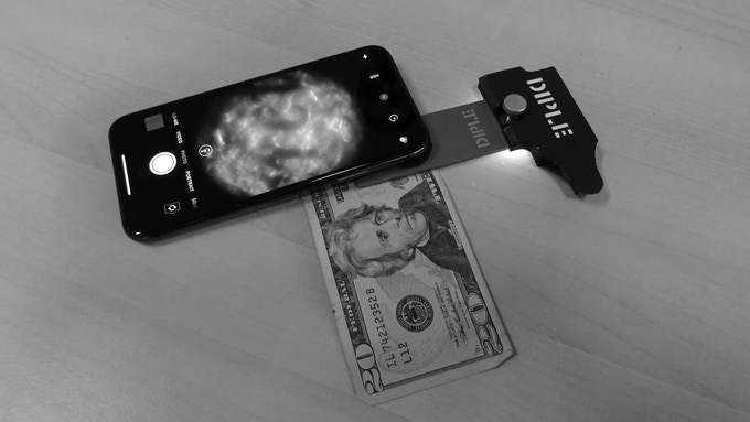
DIPLE LUX on a banknote of 20USD

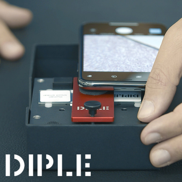
How DIPLE works
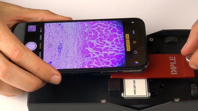
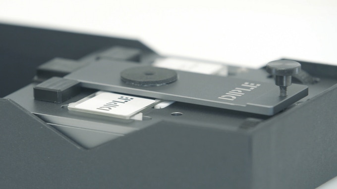
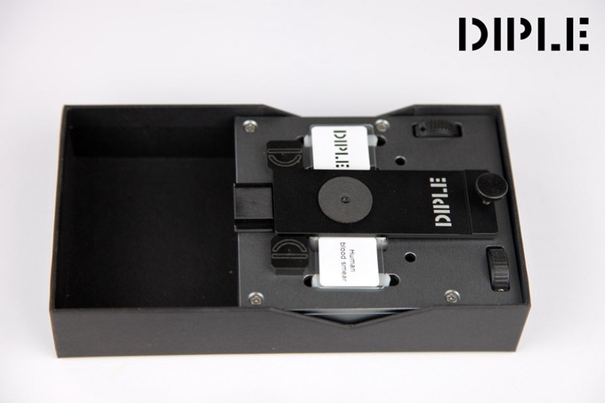
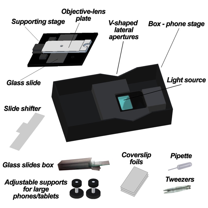
DIPLE is a compact and portable box containing a light source, a stage for samples/microscope, slides and a sturdy metal plate with an optical system.
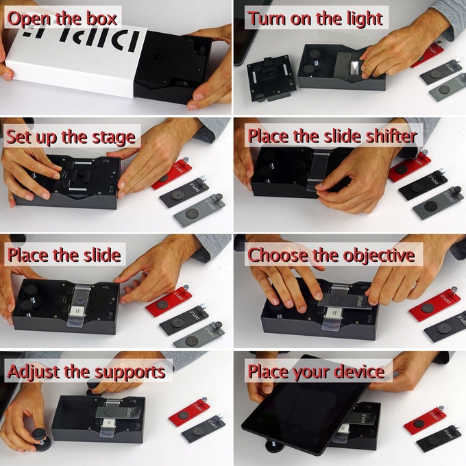
It’s straightforward to use, just lay your phone or tablet over the box, focusing the camera into the lighting hole. With the help of a magnetic guide, the objective lens and light source are automatically placed in the proper position.
Stage types
There are two different sample stages for you to pick from:
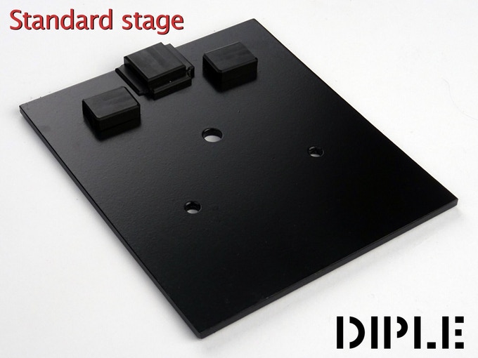
The standard stage: a simple mechanical support for your samples, where you can shift your slide manually for scanning your sample under the DIPLE lens.
OR
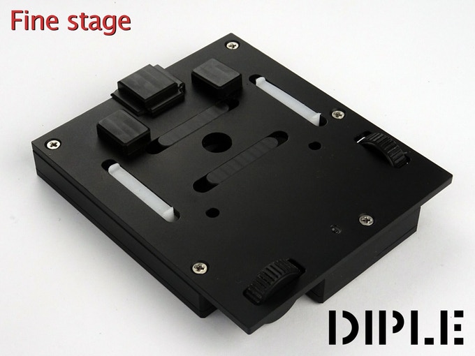
The fine stage: the slide can be shifted by mechanical screws, for a precise search of microscopic targets. The box has lateral V-shaped apertures, for allowing an easy handling of the samples, or for scanning slides over a large field.
What you can see with DIPLE
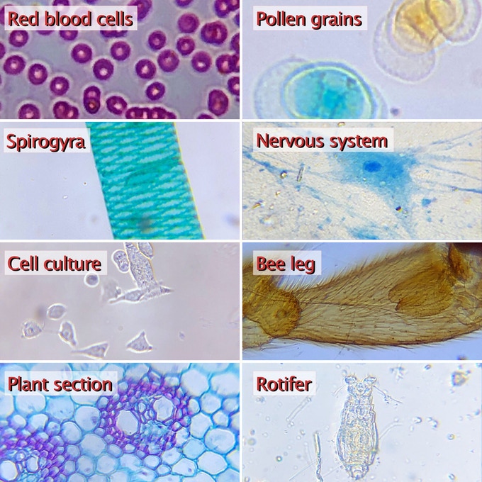
There are loads of reasons for using DIPLE, whether you’re using it for research, learning, or just to see more of the world! From the observation of microorganisms or cells to mineralogy and materials science, with DIPLE, anyone can observe the microworld in detail and save it directly to your phone, in high definition.
Here are some ideas for what you could use it for:
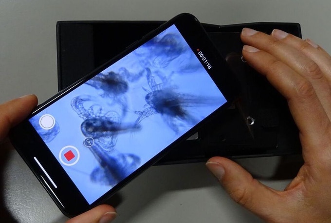
1. See your own blood cells
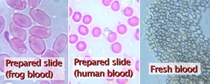
Take a tiny drop of blood and smear it on a plain glass slide. You should see only a light pink shadow. Cover it with a thin transparent plastic foil. Observe it like a standard prepared slide. You should process these cells with staining and fixing steps to get them to stay on the glass for a long time, and keep the red color. However, you are able to see the fresh cells for some minutes after their deposition even without more complex procedures.
In this short video you will see how to conduct this experiment to see your own red blood cells. In case you wanted to replicate this experiment please be careful: work in a proper environment and use only sterile lancets.
2. Micro-organisms
Collect some moss, keeping its roots in tact and with the dirt on, and place the sample in a small container, with a little water. Let it rest in the water for 1-2 days, in a warm climate. Take a drop of dirty water from the moss and put it on a glass slide. Take your 35x objective (in some cases also 75x will work fine) and lay it on the water. The objective can work in immersion, i.e. in direct contact with the water, or you can lay a plastic foil upon the liquid sample to keep it thin. Scan the sample under the objective, until you see some micro-organisms moving around, then you can zoom in to watch them in detail. When you’ve finished your observations, clean the objective lens with a wet tissue.

Rotifer filmed on DIPLE Grey
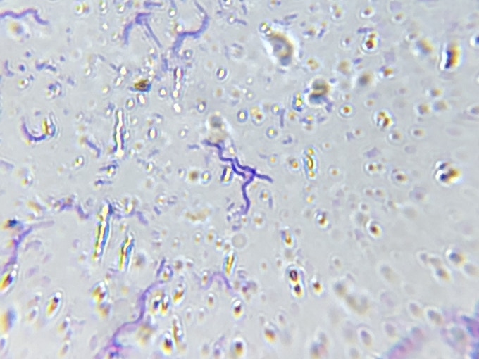
3. Bacteria
Create your own colony of bacteria by placing a mixture of nutrients (the most common is agar agar, usually mixed with meat broth) in a petri dish. Then, you can help the colonies grow by swabbing a place rich in bacteria like your mouth, your hands, or a handful of dirt and then swabbing the bacteria on the petri dish together with the nutrients. After doing this, you need to close the dish to avoid contamination and put it in a warm, safe place. After a few days (or, in some cases, even a few hours), you will get a nice collection of bacteria colonies, ready to be observed in detail.
ENGINE EXHAUST OBSERVATION
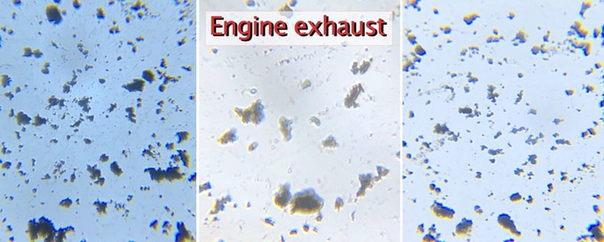
The particulate matter emitted by many car engines can be observed on DIPLE. In this video you will see this simple test, conducted by our team. We put a plain microscope slide near the exhaust tube of a car, stepped on the gas and then observed the result on our DIPLE set. The result is outstanding.
Actual magnification
Most low-budget microscopes declare false or exaggerated magnifications; see this comparison between DIPLE Red, the version which provides the lower magnification (35x REAL magnifications, multiplied by digital zoom) and a standard USB microscope (with maximum declared magnification 1000x, according to the manufacturer). In this video the two microscopes examine the same prepared slide, a botanical section. You will notice that there is a huge difference in detail, quality of the image, and definition.
HOW to use DIPLE – set up in SECONDS
NOTE: The two V-shaped openings in the box are for widening the scanning area of the sample. You can shift your sample out from the box perimetry.
DIPLE parts
Timeline
- Spring 2015 – Our first venture in microscopy begins with the creation of the initial Blips prototype
- January 2016 – The final prototype of Blips is complete
- February 2016 – SmartMicroOptics officially launches as a company
- Mid-2016 – Our Blips campaign goes live on Kickstarter
- August 2016 – The first Blips lenses are shipped to our backers
- Mid-2017 – The first DIPLE prototype is created
- First months of 2018 – The DIPLE project is developed with constant improvements on optical performance, reliability, and design
- End of 2018/first months of 2019 – The first complete prototypes are finished and sent to beta-testers
- Fall 2019 – Based on the feedback from beta-testers, the complete, fully-functional version of DIPLE, is ready to enter the global market, and our Kickstarter/Indiegogo campaigns good to go!
- January 2020 – DIPLE’s manufacturing phase begins
- May 2020 – DIPLE is ready for shipping!
Our journey so far
Following the launch of Blips, the SMO team wanted to push the magnification on mobile devices to an even higher level. In the last three years, we have worked on the development of a completely different system, based on a new design, combining very powerful micro-lenses with an aluminium cast that protects them from any damage.
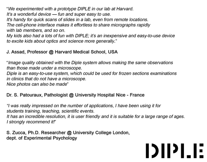
We have built prototypes, and they have been tested by scientists and researchers from top colleges and research institutions around the world, including Harvard University and the University of Cambridge.
We have also tested the durability of DIPLE. The objectives can withstand a 10-meter fall and still work perfectly! We conducted tests and launched one of our lenses from the second floor, then we used it to see red blood cells. Everything worked perfectly, as if nothing had happened! You can see the full footage of this test HERE.

*all devices with a distance photo camera-phone edge inferior to 45mm (1,77)
The Team
SmartMicroOptics S.r.l. (SMO) is a spin-off of the Italian Institute of Technology (IIT). Founded in 2016, the company made a breakthrough in the field of mobile microscopy thanks to a brand new technology, the Blips lenses & labkits, which we launched on Kickstarter in mid-2016.

Our team is young and made up of diverse personalities, with a variety of experience and skills. We’ve got the perfect combination of creativity, technical, and commercial abilities to ensure we create only high-quality products as well as deliver a reliable customer service.

ALL PICTURES AND VIDEOS OF THE MICRO-WORLD ON THIS CAMPAIGN WERE TAKEN WITH DIPLE LENSES AND COMMON SMARTPHONES
